The human body is made up of more than 37 trillion cells, self-replicating units containing microscopic structures that work together to convert nutrients into energy, among other functions. The inner mechanics of these tiny parts – called “organelles” – had long been a mystery to scientists.
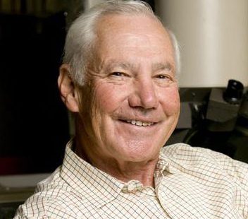
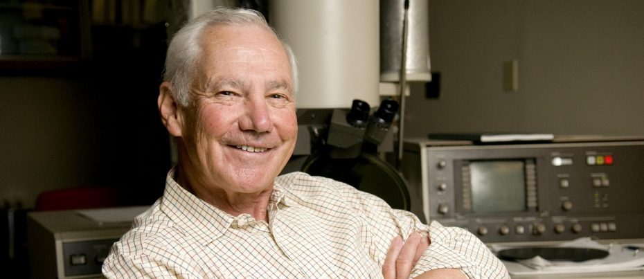
In the 1930s, Keith Roberts Porter shed light on the unknown, obtaining the first high-resolution images of individual cells using an electron microscope.
Within a layer of cells – stretched thinly using a process Porter pioneered – the team noticed a network of sacs and tubes synthesizing lipids and proteins. They named it the “endoplasmic reticulum.”
In addition, Porter helped discover skeleton-like structures called “microtubules,” which form a cell’s shape and move organelles when they are needed.
”Men have visited the moon,” he said, accepting the 1970 Horowitz Prize, “but we were the first to see particles, to see structures, that the light microscope had not been able to resolve.”